Attention: Safety Recall of Vernier Go Direct Charge Station. Click to learn more.

Shop

Human Physiology

Explore the Fundamental Principles of How the Human Body Works

Human physiology is the foundation of biomedical science and gives students the ability to understand how our bodies work. With our hands-on experiments, students collect data for measuring heart rate, EKG, blood pressure, and lung function. Whether your students are aspiring health professionals or budding biomedical engineers, they will gain a deeper understanding of human physiology through these experiments.

Featured Human Physiology Categories

We are scientists, educators, and your team

We’re here to support you as an educator as you implement data-collection technology into your teaching. See how our products provide you with affordable classroom and laboratory solutions designed for student success.

Featured Human Physiology Experiments

Introduction to Electrocardiography

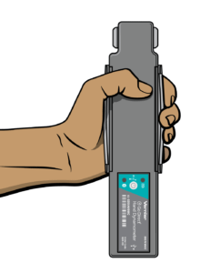
An electrocardiogram (ECG or EKG) is a graphical recording of the electrical events occurring within the heart. In a healthy heart there is a natural pacemaker in the right atrium (the sinoatrial node) which initiates an electrical sequence. This impulse then passes down natural conduction pathways between the atria to the atrioventricular node and from there to both ventricles. The natural conduction pathways facilitate orderly spread of the impulse and coordinated contraction of first the atria and then the ventricles. The electrical journey creates unique deflections in the EKG that tell a story about heart function and health. Even more information is obtained by looking at the story from different angles, which is accomplished by placing electrodes in various positions on the chest and extremities. A positive deflection in an EKG tracing represents electrical activity moving toward the active lead (the green lead in this experiment).

Five components of a single beat are traditionally recognized and labeled P, Q, R, S, and T. The P wave represents the start of the electrical journey as the impulse spreads from the sinoatrial node downward from the atria through the atrioventricular node and to the ventricles. Ventricular activation is represented by the QRS complex. The T wave results from ventricular repolarization, which is a recovery of the ventricular muscle tissue to its resting state. By looking at several beats you can also calculate the rate for each component.

Doctors and other trained personnel can look at an EKG tracing and see evidence for disorders of the heart such as abnormal slowing, speeding, irregular rhythms, injury to muscle tissue (angina), and death of muscle tissue (myocardial infarction). The length of an interval indicates whether an impulse is following its normal pathway. A long interval reveals that an impulse has been slowed or has taken a longer route. A short interval reflects an impulse which followed a shorter route. If a complex is absent, the electrical impulse did not rise normally, or was blocked at that part of the heart. Lack of normal depolarization of the atria leads to an absent P wave. An absent QRS complex after a normal P wave indicates the electrical impulse was blocked before it reached the ventricles. Abnormally shaped complexes result from abnormal spread of the impulse through the muscle tissue, such as in myocardial infarction where the impulse cannot follow its normal pathway because of tissue death or injury. Electrical patterns may also be changed by metabolic abnormalities and by various medicines.

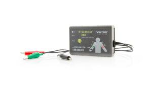
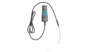
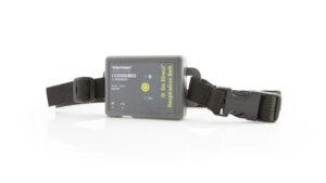
In this experiment, you will use the EKG sensor to make a five second graphical recording of your heart’s electrical activity, and then switch the red and green leads to simulate the change in electrical activity that can occur with a myocardial infarction (heart attack). You will identify the different components of the waveforms and use them to determine your heart rate. You will also determine the direction of electrical activity for the QRS complex.

Read More »

Limb Position and Grip Strength

The importance of hand strength and function is evident in all aspects of our daily living, from eating and maintaining personal hygiene to typing at the computer, performing brain surgery, or playing tennis or the piano. People suffering from arthritis or hand injury quickly appreciate the difficulty of performing even simple tasks with reduced grip strength.

Testing of hand grip strength is used by orthopedic surgeons and physical therapists to evaluate the extent of an injury and the progress of recovery. Grip strength can also be used to diagnose neuromuscular problems such as stroke, herniated disks in the neck, carpal tunnel syndrome, and elbow tendonitis. Athletes are interested in grip strength because it relates to performance in many sports, such as tennis, golf, baseball, football, gymnastics, and rock climbing.

Pinch strength is a way for occupational therapists to measure loss of fine-motor strength in the thumb, fingers, and forearm. It is useful for analyzing the extent of an injury and the outcome from surgery or therapy.

In Part I of this experiment, you will measure and compare grip strength in your right and left hands. You will also correlate grip strength with arm position, handedness, and height. In Part II you will analyze the pinch strength of each of your four fingers.

Read More »

Effect of Exercise on Heart Rate

The adaptability of the heart can be observed during exercise, when the metabolic activity of skeletal muscle tissue increases. The cardiovascular system, consisting of the heart and blood vessels, responds to exercise with an increase in heart rate and strength of contraction with each beat, resulting in a higher cardiac output (quantity of blood pumped through the heart per unit of time). Physically fit people can deliver a greater volume of blood in a single heartbeat than unfit individuals and can sustain a greater work level before reaching a maximum heart rate. Being more physically fit also leads to a more rapid recovery of resting heart rate.

In this experiment, you will observe how the heart responds to the increased metabolic demand of muscles during exercise.

Read More »

Featured Physiology Packages

SAVE/SHARE YOUR CART