Introduction
Measurement of lung volumes provides a tool for understanding normal function of the lungs as well as disease states. The breathing cycle is initiated by expansion of the chest. During quiet breathing, contraction of the diaphragm muscle causes it to flatten downward, increasing the vertical dimension of the thoracic cavity. Additional respiratory muscles in the chest (external intercostals) lift the ribs, expanding them outward and further increasing volume. The resulting increase in chest volume creates a negative pressure that draws air in through the nose and mouth. Normal exhalation is passive, resulting from “recoil” of the chest wall, diaphragm, and lung tissue. In contrast to passive breathing, active breathing uses additional muscles known as accessory respiratory muscles (including the sternocleidomastoid, scalenes, internal intercostals and abdominal muscle groups) to further expand and contract the thorax.
In normal breathing at rest, approximately one-tenth of the total lung capacity is used. Greater amounts are used as needed (e.g., with exercise). The following terms are used to describe lung volumes:
Tidal Volume (TV)
The volume of air breathed in and out without conscious effort
Inspiratory Reserve Volume (IRV)
The additional volume of air that can be inhaled with maximum effort after a normal inspiration
Expiratory Reserve Volume (ERV)
The additional volume of air that can be forcibly exhaled after normal exhalation
Vital Capacity (VC)
The total volume of air that can be exhaled after a maximum inhalation:

Residual Volume (RV)
The volume of air remaining in the lungs after maximum exhalation (the lungs can never be completely emptied)
Total Lung Capacity (TLC)

Minute Ventilation
The volume of air breathed in 1 minute:
")
In this experiment, you will measure lung volumes during normal breathing and with maximum effort. You will correlate lung volumes with a variety of clinical scenarios.
Important: The equipment used in this experiment is for educational purposes only and should not be used to diagnose medical conditions.
Objectives
- Obtain graphical representation of lung capacities and volumes.
- Compare lung volumes between males and females.
- Correlate lung volumes with clinical conditions.
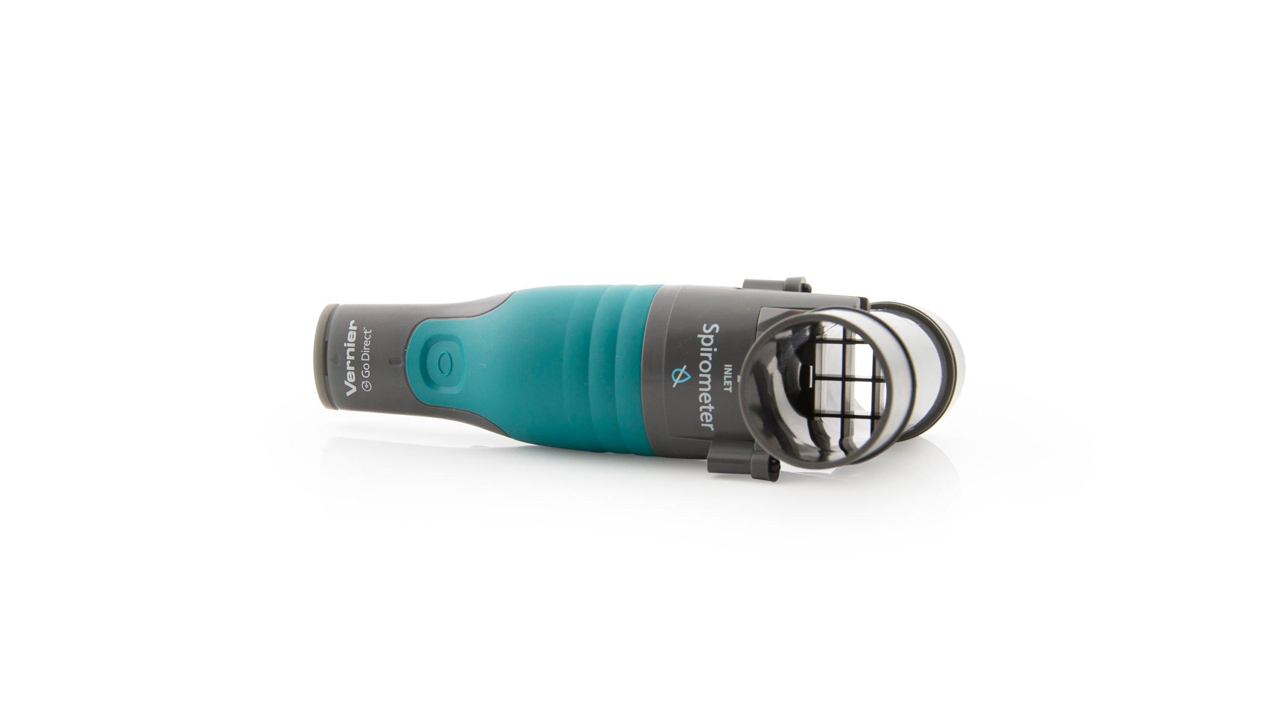
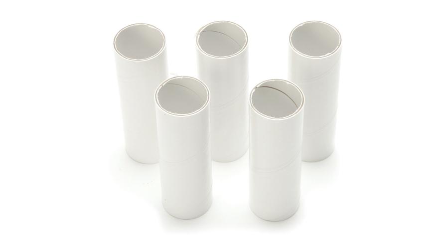
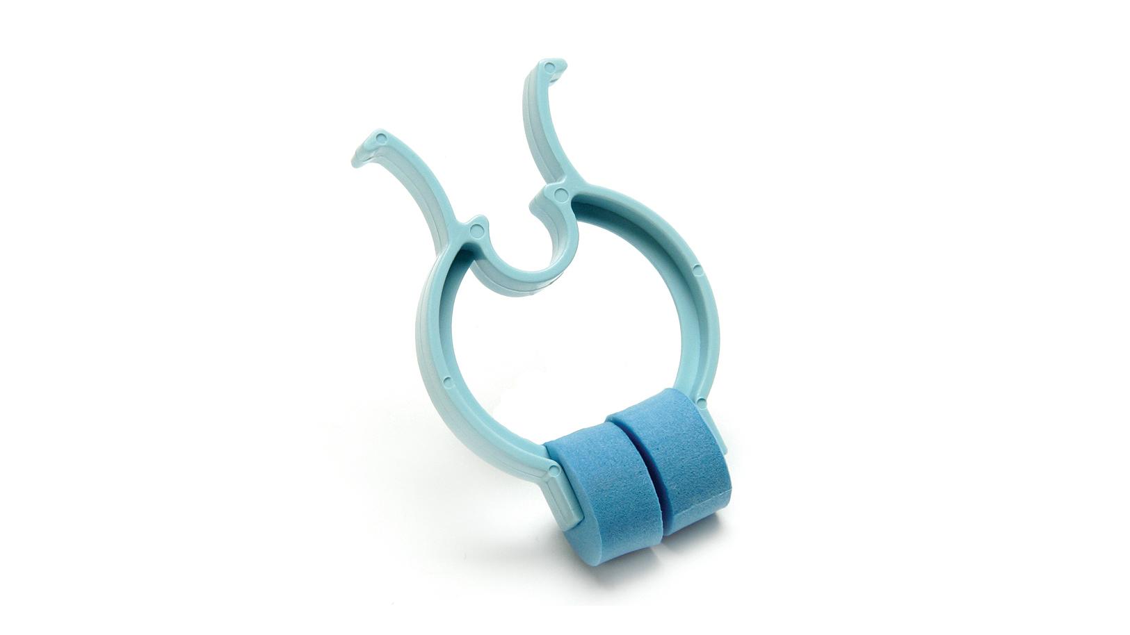
Sensors and Equipment
This experiment features the following sensors and equipment. Additional equipment may be required.

Correlations
Teaching to an educational standard? This experiment supports the standards below.
- International Baccalaureate (IB)/Sports, Exercise, and Health Science
- 2.1 Structure and function of the ventilatory system
- International Baccalaureate (IB) 2025/Biology
- B3.1.6—Measurement of lung volumes
Ready to Experiment?
Ask an Expert
Get answers to your questions about how to teach this experiment with our support team.
- Call toll-free: 888-837-6437
- Chat with Us
- Email support@vernier.com
Purchase the Lab Book
This experiment is #7 of Human Physiology Experiments: Volume 2. The experiment in the book includes student instructions as well as instructor information for set up, helpful hints, and sample graphs and data.