Introduction
Electrical activity of the heart is closely associated with pressure changes in the heart chambers and blood vessels. Understanding the events of the cardiac conduction system, the muscle activity of the heart and the pumping events that follow, is important for understanding how blood pressure is maintained during the cardiac cycle.
An electrocardiogram (ECG or EKG) is a graphical recording of the electrical events occurring within the heart. In a healthy heart, a natural pacemaker in the right atrium (the sinoatrial node) initiates an electrical sequence. This impulse passes down natural conduction pathways from the atria to the atrioventricular node and from there continues down the right and left bundle branches to both ventricles. The natural conduction pathways facilitate orderly spread of the impulse and coordinated contraction of first the atria and then the ventricles. Five components of a single EKG are traditionally recognized and labeled P, Q, R, S, and T (see Figure 1). The P wave represents the start of the electrical journey as the impulse spreads from the sinoatrial node downward from the atria through the atrioventricular node and to the ventricles. Ventricular activation is represented by the QRS complex, which initiates contraction of the ventricles. The T wave results from ventricular repolarization, which is a recovery of the ventricular muscle tissue to its resting state.
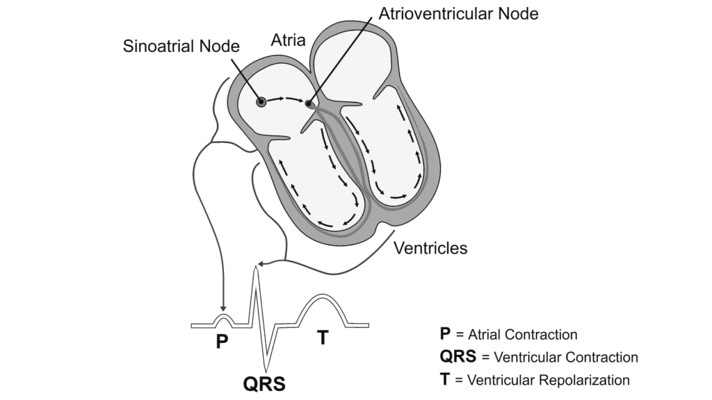
Figure 1
Blood pressure is a measure of the changing fluid pressure within the circulatory system. It varies from a peak pressure produced by contraction of the left ventricle, to a low pressure, which is maintained by closure of the aortic valve and elastic recoil of the arterial system. The peak pressure is called systole, and the pressure that is maintained even while the left ventricle is relaxing is called diastole.
If you could measure arterial blood pressure from an artery while you performed an EKG, you would see that each QRS complex precedes a pressure pulse in the artery. The time that it takes this pressure pulse to travel from the ventricles to the artery is referred to as the Pulse Transit Time. Because the ventricular contraction and the QRS complex happen almost simultaneously, you can use the peak of the R wave as an indicator of the time ventricular contraction occurred. Using a blood pressure cuff on the arm, you will be able to record the following pressure pulses in the brachial artery. By recording these events, you will be able to calculate the Pulse Transit Time.
Important: The equipment used in this experiment is for educational purposes only and should not be used to diagnose medical conditions.
Objectives
- Obtain graphical representation of the electrical activity of the heart (EKG).
- Obtain graphical representation of a pressure pulse from the brachial artery.
- Calculate the transit time of a pressure pulse from the heart to the brachial artery.
Sensors and Equipment
This experiment features the following sensors and equipment. Additional equipment may be required.


Correlations
Teaching to an educational standard? This experiment supports the standards below.
- International Baccalaureate (IB) 2025/Biology
- B3.2.16—Stages in the cardiac cycle
Ready to Experiment?
Ask an Expert
Get answers to your questions about how to teach this experiment with our support team.
- Call toll-free: 888-837-6437
- Chat with Us
- Email support@vernier.com
Purchase the Lab Book
This experiment is #4 of Human Physiology Experiments: Volume 2. The experiment in the book includes student instructions as well as instructor information for set up, helpful hints, and sample graphs and data.